Alzheimer’s Disease: a brain disorder that usually starts in late middle age or old age and gets worse over time. Symptoms include loss of memory, confusion, difficulty thinking, and changes in language, behavior, and personality. (NCI3) A “degenerative” disease whose fundamental cause is dying and damaged cells. (Ratey, 49) Characterized by deficits in recent memory. It results from the loss of “synapses.” The brain can regrow synapses in the early stages of the disease, but in the later stages, neurons actually die. Our brain cannot regrow neurons, so this cell death results in permanent damage. (Kandel4, 117) First appears as progressive "memory" loss and later develops into generalized “dementia.” (Kolb, 170) "Beta-amyloid plaques" and tangles of proteins inside neurons are characteristic features of brain tissue in patients with Alzheimer’s. (Fields, 311)
The discovery of Alzheimer’s disease dates back to 1906, when Alois Alzheimer described the case of a fifty-one year old woman who had become suddenly and irrationally jealous of her husband. She died less than five years after the onset of her symptoms. Alzheimer performed an autopsy on her and found three specific alternations in the cerebral cortex. First, her brain was shrunken and atrophied. Second, the outside of the nerve cells contained deposits of a dense material that formed what we now call amyloid plaques. Third, inside the neurons was an accumulation of tangled protein fibers that we now call “neurofibrillary tangles.” (Kandel4, 118) Relentlessly robs its sufferers of their memories and "personality." 4.5 million Americans have this “neurodegenerative” disorder. Found in nearly half of those older than 85. Preventing Alzheimer’s at an early stage may be the only hope for those at risk. Marijuana may stall the disease. (SAM April/May 2007, 14) The most significant “risk factor” found to date is the APOE gene. This gene codes for a protein that combines with fats to form a class of molecules called lipoproteins. One variation of this gene is APOE4. About half of people with… Alzheimer’s have this (variation). A number of studies have shown that “type 2 diabetes” is a risk factor for Alzheimer’s disease. Environmental factors… may also contribute to the susceptibility to Alzheimer’s disease, but all studies to date point to amyloid clumping as the fundamental cause of dementia. Recent studies have therefore focused on preventing clumping and clearing preexisting amyloid clumps by using “antibodies” that specifically recognize these clumps. (Kandel4, 124-125) One Alzheimer’s study showed that our chances for developing Alzheimer’s drops 17% for every year of education we have beyond high school. (Ratey, 231) Some of the most disturbing changes caused by Alzheimer’s disease occur when blood flow, activity, and neuronal connections in the “association cortex” are compromised. (Bainbridge, 264) In the early stages of Alzheimer’s, the symptoms of memory impairment are often mild, such as forgetting the names of familiar people, forgetting the location of familiar places, or forgetting to do things. As the disease progresses, memory loss and confusion become more pervasive. The person becomes unable to remember what month it is or the names of family members. Frustrated and disoriented by the inability to retrieve even simple information, the person can become agitated and moody. In the last stage, brain damage is widespread. The person no longer recognizes loved ones and is unable to communicate in any meaningful way. (Hockenbury, 252) At first, the brain is able to compensate well enough that even a family member can’t tell the difference between someone who has this initial damage and someone who does not. Over time, however, as more and more connections are damaged and neurons begin to die, regions like the “hippocampus” disintegrate and the brain begins to lose crucial functions such as memory storage. Symptoms related to memory loss then become noticeable. Treatment for Alzheimer’s is likely to be most effective early on, before extensive cell death, so “neurologists” are trying to develop functional brain imaging and other methods of identifying the disease as early as possible. (Kandel4, 117-119)
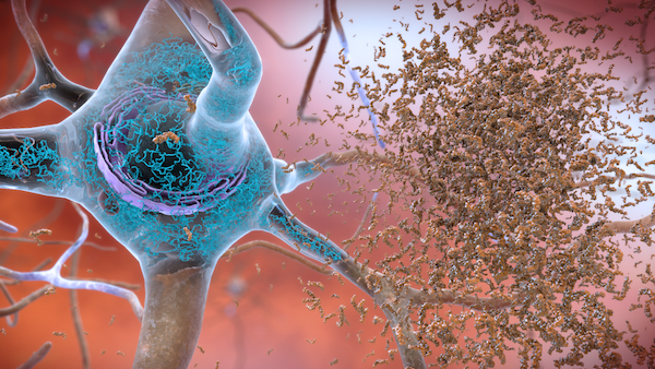
Beta-Amyloid Plaques: dense deposits of protein and other cell materials outside and around neurons. Found in abundance in Alzheimer’s patients. (Hockenbury, 252) Clumps of the abnormal proteins. Build up in certain organs. This reduces their ability to work correctly. (PubMedHealth2) Plaques initially form in specific, restricted areas on the brain. One such site is the “prefrontal cortex,” the part of the brain involved in attention, self-control, and problem solving. Amyloid-beta peptide is responsible for forming amyloid plaques. This peptide is a part of a much larger protein called the ‘amyloid precursor protein’ which is thought to be lodged in the cell membrane of “dendrites.” Two separate “enzymes” cut through the precursor protein, each in a different place, releasing the amyloid-beta peptide. Once released from the cell membrane, the peptide floats in the space outside the neuron. The production and liberation of the amyloid-beta peptide are normal occurrences in everyone’s brain. In people with Alzheimer’s disease, production of the protein may be accelerated, or clearance of the protein from the area surrounding the cell may be slowed. Either action can result in abnormal accumulations of peptides. What’s more, these peptides are sticky. They adhere to one another and ultimately form the amyloid plaques. (Kandel4, 118-121) Proteins deposited in the brain, forming plaques that cause signaling problems between neurons, triggering inflammation within the brain. (Leschziner, 86) A … form of ‘amyloid beta-peptides’ is the major component of amyloid plaques found in individuals with Alzheimer's disease and in aged individuals with “Down syndrome.” The peptide is found predominantly in the "nervous system," but there have been reports of its presence in non-neural tissue. (MeSH) Alzheimer’s signature beta-amyloid crystals and micro-fibrillary tangles… are easily revealed in brain images in about half of normal 70-year olds. Yet, by their 70th birthdays, only 7% of people have received a formal Alzheimer’s or related “senile dementia” diagnosis. Sadly, the pathological seeds of greater troubles have already been planted in the majority of their 70-year old brains. (Merzenich, 5) They form ten to fifteen years before a person’s memory or thinking has begun to change. If these structures could be detected when they first appear, it might be possible to prevent damage to the brain and to stop Alzheimer’s disease in its tracks. (Kandel4, 118) In humans, levels of beta-amyloid in the “cerebrospinal fluid,” where the fluid inside the “glymphatic system” vessels end up, are highest in the morning, suggesting a similar flushing-out of beta-amyloid overnight. A recent study in humans has shown that even after a single night of “sleep deprivation,” levels of beta-amyloid in certain parts of the brain, including the hippocampus, often damaged in Alzheimer’s, go up. (Leschziner, 87)
Inherited Alzheimer’s: a rare, early-onset form (that) runs strongly in some families. While Alzheimer’s disease usually occurs in people in their seventies or eighties whose families have no history of the disease, (Inherited Alzheimer’s) starts with a mutation in the gene for the amyloid precursor protein that causes the peptides to clump. Families with inherited Alzheimer’s have mutations in the genes that code for a protein called ‘presenilin.’ These mutations prevent presenilin from helping to digest amyloid-beta peptides floating in the space between neurons. (Kandel4, 122-124) Most cases of early-onset Alzheimer’s disease are caused by gene mutations that can be passed from parent to child. Researchers have found that this form of the disorder can result from mutations in one of three genes: ‘APP,’ ‘PSEN1,’ or ‘PSEN2.’ When any of these genes is altered, large amounts of a toxic protein fragment called ‘amyloid beta peptide’ are produced in the brain. This peptide can build up in the brain to form clumps (of) amyloid plaques. A buildup of toxic amyloid beta peptide and amyloid plaques may lead to the death of nerve cells and the progressive signs and symptoms of this disorder. (GHR)
Neurofibrillary Tangles: twisted fibers that build up inside the neuron. Found to a great extent in Alzheimer’s patients. (Hockenbury, 252) Tangles start in the hippocampus. (Kandel4, 118) A “pathological” accumulation of paired (protein fibers) composed of abnormally-formed ‘tau protein’ that is found chiefly in the “cytoplasm” of nerve cells of the brain and especially the “cerebral cortex” and hippocampus. (GHR) Abnormal structures located in various parts of the brain and composed of dense arrays of paired “neurofilaments” and “microtubules.” As one of the hallmarks of Alzheimer’s disease, the neurofibrillary tangles eventually occupy the whole of the cytoplasm in certain classes of cell in the cerebral cortex, hippocampus, “brain stem,” and “diencephalon.” The number of these tangles correlates with the degree of dementia during life. Some studies suggest that tangle “antigens” leak into the “circulation” both in the course of normal aging and in cases of Alzheimer’s disease. (MeSH) Tau (protein) is located inside the neuron. When a molecular defect causes the tau protein to misfold, it forms toxic clumps that create neurofibrillary tangles. (Kandel4, 121)